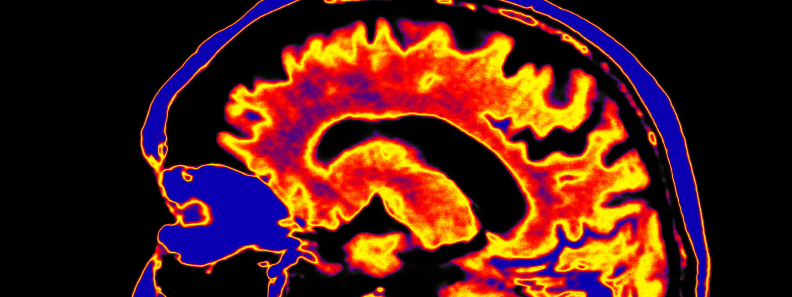
A system using light for non-invasive monitoring of Alzheimer’s Disease is to be explored in research led at the University of Strathclyde.
The system will use single-pixel imaging to measure activity in the brain, and the researchers aim to complement existing technology by enhancing its fidelity while also making the process safer and less expensive.
A technique currently used in brain imaging, known as diffuse optical tomography (DOT), works well in observing the brain’s surface activity but has less capacity to measure electrical or deep tissue activity.
The new research will investigate whether single-pixel imaging techniques could help augment DOT by capturing volumetric imaging, a type of imaging which DOT is unable to produce.
Dr Akhil Kallepalli, a Chancellor’s Fellow in Strathclyde’s Department of Biomedical Engineering and a Leverhulme Early Career Fellow, will work on developing this combination of techniques with a team from Washington University in St. Louis, Missouri, led by Professor Joseph Culver.
Wearable headset
Dr Kallepalli said: “The DOT technique examines the brain with the use of a wearable headset with several optical diodes. As there are types of brain activity which DOT does not measure, my proposal is to develop another technique which is capable of doing so.
“With single-pixel imaging, you project light and record the returned optical signal after it interacts with an object. This measurement is made with a ‘bucket’ detector such as a photomultiplier tube or single-photon avalanche diode (SPAD); devices which measure an electrical value. We want to find out if we can use this in a DOT-compatible configuration to increase the fidelity of DOT for deep tissue imaging and to combine two separate techniques to look at the brain differently.
“My interest is in finding out where we can look deeper into the brain tissue so that we can try and understand the haemodynamics of Alzheimer's Disease. Using light is safer and less expensive than many more complex techniques for exploring Alzheimer’s, which is generalized to an abnormal build-up of proteins, called amyloid and tau and calcium in the human brain. We are at the early stages of developing this technique, but we believe it is highly promising.”
Dr Kallepalli will work on the project at Professor Culver’s laboratory at Washington University School of Medicine’s Mallinckrodt Institute of Radiology, which contains a range of high-density DOT scanners. Researchers from Imperial College London will also participate in the project, where Dr Kallepalli holds an honorary visiting position.
Dr Kallepalli has received an EPSRC Overseas Travel Grant to support the establishment of the collaboration. He began the development of the project idea while working at the University of Glasgow.