Working with Steve Blair's team at the University of Utah, we have developed an optical analogue of the well-known Utah Electrode Array. This Utah Optrode Array (UOA) has been developed for optogenetic experiments. The device allows for precise multi-site light delivery in deep cortical layers. It consists of an array of 100 glass microneedles paired with a µLED array and is capable of optogenetically exciting thousands of neurons in vivo in spatiotemporal patterns. As the device is electrically controlled, it is compact and does not require an external optical system or to be tethered to bulky fibre optics.
NeurophotonicsUtah Optrode Array
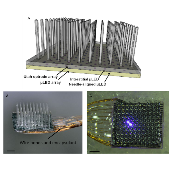
The Utah Optrode Array
A Schematic of the device design including the glass needle array and bonded µLED array. Light from the µLEDs is collimated by either a metal pinhole structure or microfabricated silicon optical interposer. Light is guided down the needles and emitted from the tips of the needles. Interstitial µLEDs provide cortical surface illumination. B Completed device, including wire bonds and encapsulant. C Close-up showing a single µLED (80µm in size) illuminated. Scale bars are 1 mm.
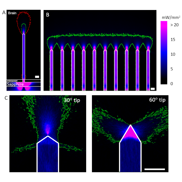
Optical Modelling
Optical modelling is used extensively to give an accurate picture of the light distribution in brain tissue. A Cross-section of the modelled light output from the Utah Optrode Array. The optical interposer collimates the emission from the µLED and greatly reduces stray light. B Cross section when a row of LEDs is illuminated simultaneously. C The tip shape has a significant effect on the light distribution in tissue. In all images the green contour marks a 1 mW/mm2 irradiance boundary, while the red line indicates the 0.1mW/mm2 level. Scale bars are 100 µm.
Our Publications


Funding for this work was provided by the NIH BRAIN Initiative Program, through Grant No. U01 NS099702. Additional support comes from the Royal Academy of Engineering through their Chair in Emerging Technology scheme.